

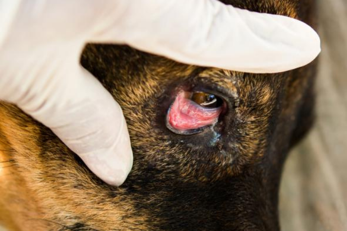
“CHERRY EYE” or PROTRUSION OF THE GLAND OF THE 3rd EYELID
Protrusion of the gland of the third eyelid (or “cherry eye”) occurs most commonly in dogs and occasionally in cats. The appearance is characteristic, with the gland of the third eyelid protruding as a reddish follicular mass from behind a usually “floppy” margin of the third eyelid (Figure 1). The gland should be surgically replaced to retain essential lacrimal function and to prevent the exposed gland and overlying conjunctiva from becoming dry, inflamed, secondarily infected, and cosmetically unappealing. Prolapsed glands of the 3rd eyelid should never be removed because the gland of the third eyelid is a significant contributor to precorneal tear film production. Studies confirm clinical experience that keratoconjunctivitis sicca (KCS) is commonly seen, often years later, in animals, especially those of susceptible breeds in which the 3rd eyelid or its gland was removed. Also, complications have been reported in prolapsed glands left in the prolapsed position.
Treatment
Occasionally in the early stages, a prolapsed gland can be manipulated into its normal position. However, recurrence is almost inevitable. For these reasons, surgical replacement is practiced. If the gland is severely inflamed or the conjunctival surface secondarily infected, preoperative treatment for a few days with a topical antibiotic-steroid ointment is advisable; however, this treatment will not result in resolution of protrusion.
The most common technique performed is the “pocket technique” (Figure 2), where the gland is “buried” back into the third eyelid.
Postoperative medical treatment includes topical antibiotic-steroid ointment or solution and the use of an Elizabethan collar. Oral administration of a nonsteroidal anti-inflammatory agent may be indicated for postoperative analgesia. Recurrence is possible, even with a correctly performed pocket or anchoring technique, especially in the very large breed dogs such as mastiffs and Newfoundlands.
Prolapsed glands of the third eyelid are treated by replacement, not by excision.
recent post
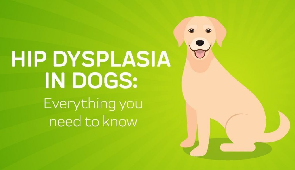
EARLY DETECTION AND TREATMENT OF HIP DYSPLASIA IN PUPPIES
Hip Dysplasia in dogs is an anatomical orthopedic condition which has a genetic background and affects mostly..
learn more
“CHERRY EYE” or PROTRUSION OF THE GLAND OF THE 3rd EYELID
Protrusion of the gland of the third eyelid (or “cherry eye”) occurs most commonly in dogs and..
learn more
